Table of Contents
Gnathology is the branch of Dentistry that deals with dental occlusion, malocclusions and the so-called cranio or temporomandibular disorders. In other words, it is the Dental Specialty that studies and treats those dysfunctions and pathologies of the masticatory system of functional and not strictly dental origin.
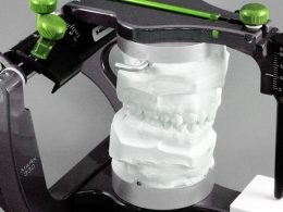
Dental occlusion is defined as the way the teeth contact each other when the lower jaw (mandible) and the upper jaw (maxillary bone) come together or relate. Refers to the gear of the teeth in any kind of functional relationship. The “normal” occlusion is always desirable, since it is the one that allows oral functions to perform correctly, provides the best facial aesthetics and is essential for the prevention of many diseases.
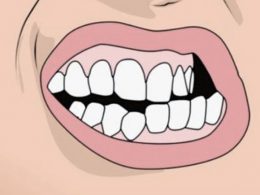
As long as there is no normal occlusion in the mouth, we speak of malocclusion. Malocclusion can affect teeth, speech, aesthetics (dental and facial) and other functions of the oral cavity; mainly the masticatory function. Malocclusion can occur from a wide variety of factors including, among others; genetic inheritance, eruption problems, loss of teeth that are not replaced later, trauma, disease, bad habits and poor dental practices.
Gnathology, in fact, focuses exclusively on the muscles of mastication and the only mobile joint of the entire head; the temporomandibular joint (TMJ), which is the joint that forms between the lower jaw or mandible and the skull.
The real disorders and dysfunctions of these anatomical structures, many times caused by malocclusions, are very common, although they generally go unnoticed because they are not always symptomatic. Unfortunately, it must be said that many Dentists only have a vague knowledge about Gnathology, and many times based on irrational, erroneous or outdated concepts.
For this reason, the diagnosis is often vague, difficult and late; the therapeutic approach confusing, and the clinical results quite limited. At DENTAL VIP we always try to keep up to date with the advances and new concepts of this Specialty, through congresses and continuing education courses, which allow us to address with greater precision and knowledge one of the darkest and most ambiguous fields of contemporary Dentistry; dental occlusion.
Chewing or Masticatory Function
Chewing is the first part of the digestive function. It is the process by which we grind food in the oral cavity.
When putting food in the mouth and chewing, a salivary secretion occurs due to a congenital reflex action. Saliva is a complex organic fluid produced by the salivary glands of the oral cavity, and involved in the first phase of digestion.
During chewing, the teeth, lips, jaws, temporomandibular joint, cheeks, tongue and hard palate (the latter passively); work in a jointly and coordinated way.
The maxillary bones and the temporomandibular joint, in addition to having the ability to close with remarkable force, also perform lateral movements that help to grind the food more and more finely.
In summary we can say that chewing is the action of breaking down foods, preparatory to the act of swallowing. This decomposition action is a complex set of highly organized neuromuscular and endocrine activities, very necessary for the correct feeding process.
The objective of chewing is to triturate, grind and mix the foods with saliva, so that they can be introduced by swallowing into the digestive canal.
Components of the Masticatory System
The masticatory system is a functional unit composed of the teeth and their supporting structures, the jaws, the temporomandibular joints, the muscles directly and indirectly involved in mastication (including the muscles of the lips and tongue) and the vessels and nerves that irrigate and innervate all these tissues.
Chewing Muscles
The masticatory muscles (or chewing muscles) are responsible for the movements of the lower jaw when the person eats or feeds. The chewing muscles originate in the skull and insert into the mandible, thus acting on chewing and other movements of the bone.
Exist mainly four masticatory muscles on each side of the head: masseter, temporal, external or lateral pterygoid and internal or medial pterygoid.
Temporomandibular Joint (TMJ)
In anatomy, the temporomandibular joints (TMJs) are the two joints that connect the lower jaw to the skull. It is a bilateral synovial joint between the temporal bone of the skull above and the mandible below; and it is from these bones that it derives its name.
This joint is unique throughout the body because it is a bilateral joint that functions as a single unit. Since the TMJ is connected to the lower jaw, the right and left joints must work together, and therefore, they are not independent of each other.
Its main components are the joint capsule, the articular disc or meniscus, the mandibular condyles, the articular surfaces of the temporal bone (fossa and tubercle), the temporomandibular ligament, the stylomandibular ligament and the sphenomandibular ligament.
Physiology of Mastication
During mastication, only the lower jaw can move, since the upper jaw is fixed and for functional purposes, it is part of the skull.
The teeth determine the movement pattern and positions of the mandible when a person chews, and therefore, also determine the positions of the mandibular condyles within the TMJs. Therefore, the functioning of the joints and the chewing muscles is directed and determined by the teeth and the way in which they are related to each other (dental occlusion).
As the muscles contract, the jaw has two heads or articular surfaces called “condyles”, which move along two cavities housed in the base of the skull (mandibular fossae or glenoid cavities of the temporal bones).
Between these two bone structures (condyle and cavity) there is a cartilage disc that acts as a cushion or shock absorber to prevent the bones from wearing down by friction. The disc is located inside an articular capsule and is located and displaced by the action of various ligaments.
When the mouth is opened, the condyles slide forward and downward, following the glenoid fossa and, at maximum opening, almost reach the upper portion of the articular tubercle. The discs accompany the condyles in this movement, remaining constantly on them thanks to their ligaments. Similarly, the mandible performs its lateral movements, always with the articular menisci accompanying the condyles.
Origin of Gnathological Problems
Muscles are made up of bundles of muscle fibers, and these fibers have a certain resting length at which they function best, their “physiological rest position”. At this length, the muscle is in its strongest, most stable, and most relaxed position.
If due to the bad position of the teeth or jaws (malocclusions), bad habits (bruxism), facial trauma or defective dental restorations (high fillings, crowns or bridges) the muscles must always be stretched or contracted excessively to “accommodate the mandible” when chewing; constant tension causes blood circulation to decrease, muscle tone increases, spasms occur, ligament function is affected and the correct functioning of the articular meniscus is altered (accompany the mandibular condyle in all its movements).
The constant muscular hypertonicity, in a short time, will cause the articular discs to begin to advance or delay; then allowing friction wear to occur on the bony surfaces of the joints, that is, TMJ pathologies or temporomandibular disorders.
Although masticatory dysfunction is the main cause of TMJ disorders, they can also be the result of trauma, excessive emotional stress, arthritis, infections, degenerative collagen diseases, or tumors in the area.
Consequences of TMJ Disorders
If you experience pain, clicking, or discomfort in your jaw, it may be an indication of a TMJ disorder. This condition affects a large percentage of the population, yet the number of people receiving treatment for symptoms represents only a small fraction.
Since TMJ symptoms are late-onset, small discomforts at the joint level are usually ignored. When left untreated, they can become chronic, painful and can affect the normal development of the person.
Disharmonies between the relationship of the teeth, the masticatory muscles and temporomandibular joints can cause:
- Migraine and Facial Pain Muscle hypertonicity associated with occlusal disease favors the accumulation of large amounts of lactic acid in the masticatory muscles, which is responsible for facial pain. In addition, the pain can easily refer to neighboring areas and trigger migraines and headaches, many times classified as idiopathic by Doctors.
- Discomfort in the Back, Neck and Shoulders Problems of this type affect not only the face and head area, but also other areas of the body. Muscle fatigue usually begins in the lower jaw, but the pain can radiate to the neck, vertebrae, shoulders, back; and even, to the hips.
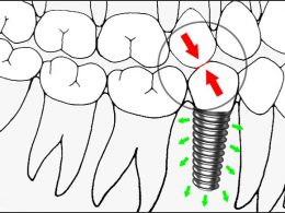
- Dental Wear Wear facets are flat and polished areas seen on the biting surfaces (occlusal faces or incisal edges) of teeth, usually caused by premature contacts, occlusal interferences, or parafunctional habits. Wear is caused by excessive chewing force or by poorly distributed or functionally aberrant forces.
- Dental Pain and Sensitivity Having occlusal trauma, abfractions (small wears and fractures) occur at the level of the neck of the teeth, leaving exposed permeable dentin. In addition, when tooth wear is very severe, it can reach the dental pulp; causing sensitivity, acute or chronic dental pain and other typical symptoms of pulp necrosis.
- Tooth Mobility Although mobility is one of the most characteristic signs of periodontal disease, it can also be the consequence of occlusal trauma due to mandibular dysfunction. When a tooth receives a greater load than it is physiologically capable of supporting, its periodontal ligament becomes inflamed and its mechanical stability decreases.
- Coronal Chipping and Fractures Over time, the accumulation of excessive forces considerably wears down enamel and dentin, creating cracks in the mineralized tissue of the teeth and making them susceptible to large fractures. The same is true for porcelain restorations, which collapse and lose their structural integrity due to excessive functional or parafunctional stress.
- Edentulism As the dysfunction becomes chronic, the sum of tooth wear and fractures leads to a point of no return, where teeth cannot be restored due to a lack of healthy tooth structure. In these cases, the extraction of all the remaining roots becomes pertinent to avoid infections of pulp etiology.
- Damage and Hearing Loss Since the lower jaw connects to the skull near the location of the ear canal, inflammation of the muscles and structures in the area can have reaching effects. The area around the ear canal is sensitive and can be affected by the symptoms of TMJ disorders over time. Severe cases can cause tinnitus (ringing in the ears) and other damage that ultimately leads to partial hearing loss.
- Functional Disability In some cases, the evolution of the disease can be so aggressive that it completely destroys the temporomandibular joints, permanently prevents any oral movement and requires the application of complex reconstructive techniques of Maxillofacial Surgery (with autologous bone grafts) to “build new joints” and restoring the patient’s chewing function.
Treatment of Masticatory Dysfunction
The development of a treatment plan for a patient with temporomandibular joint syndrome should be based on a comprehensive study of the patient’s mandibular, cranial, cervical and general health status. Also, sometimes it is necessary to carry out complementary tests and studies such as blood tests, X-rays and magnetic resonances; to name just a few.
It is important to mention that therapy for these types of problems is not easy, and that a single treatment modality is rarely enough to overcome this type of pathologies.
In the most complex cases, collaboration between various Specialists (Dentist, Physical Therapist, Psychologist, Otolaryngologist, Neurologist and the Maxillofacial Surgeon occasionally) it is essential to be able to offer the patient an effective and individualized treatment plan for his needs. If you want to know more about it, the following publication is for you: Management and Treatment of Temporomandibular Disorders.
“Dental Occlusion, Temporomandibular Joint and Masticatory Function, Are the 3 Basic Concepts Associated With the Specialty of Gnathology”.
DENTAL TIP
Save Up to 70% on Your Dental Quote!
Never forget that dental tourism is an extraordinary tool for accessing top-notch Dentistry.
DENTAL VIP trusts that quality of dental care should be affordable to all, no matter where you live. Hundreds of patients come to Venezuela for state-of-the-art medical and dental care every year, saving large amounts of money on health. Our rates are significantly lower than in any other Latin American country, without it implying a decrease or compromise in the quality of service. Simply what happens is that property costs and salary levels are both much lower than in any other place, which translates in better offers for our patients.
We guarantee that by using the services offered by DENTAL VIP, you could save up to 70% on your dental quote, compared to the local rates stipulated in your country.
Come for the price and stay for the quality! Contact us today via WhatsApp +58 414-9033547 or Email and overcome once and for all any barrier that prevents you from show off a white, healthy and beautiful smile.
