
Table of Contents
There are many reasons to use dental photography today, however, the main objective of digital photography in dental tourism is to know with precision the clinical conditions of the patient’s oral cavity to diagnose, plan treatments and prepare budgets.
Its other uses include legal case documentation, publication, education, communication with patients, professional staff members, colleagues and dental technicians and; finally, marketing. Each of these uses improves and raises the quality of dental practice and services delivery to patients.
Therefore, dental photography should be considered an essential diagnostic tool, just like X-rays and other complementary tests. It allows us to observe and analyze the facial profile, the characteristics of the smile, the degree of exposure of the gingiva at rest and natural smile, the number of teeth present and absent, dental alignment, the presence of cavities and occlusal disharmonies, prosthetic needs, the state of gingival health and periodontal involvement; and even, the morphology of the residual ridge before the placement of dental implants.
Digital images constitute an invaluable visual record and offer us the possibility of monitoring particular clinical situations for long periods of time.
Can I Take the Photographs Myself?
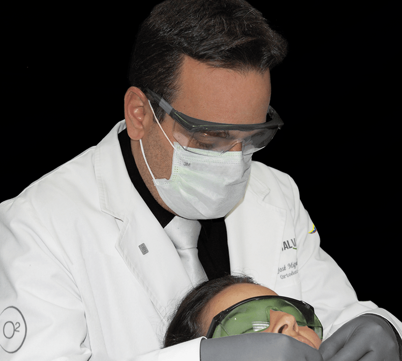
In reality, it is very difficult, since to achieve precise images, in addition to the pertinent photographic skills; it is necessary to use a series of resources typical of Dentistry. Ring flashes, cheek retractors and intraoral mirrors are essential elements of professional dental photography.
Likewise, it is essential to know the protocol of the projections that are usually required for study and analysis purposes. Recording of details is important for examination, diagnosis, treatment planning, and evaluation of clinical outcomes in many disciplines; including smile design, implantology, oral surgery, orthodontics, and prosthodontics.
The best thing to do is to go to an oral diagnostic center in your area so that a truly trained technician can perform the study.
What Requirements Must the Images Meet?
Basically high resolution, good lighting, sharpness and depth of field.
Other important elements are all those related to color reproduction (tone, hue, saturation, temperature and white balance), since it is necessary for any dental image to accurately register and reproduce the color that the human eye perceives at the time of visual inspection.
Correct color rendering is an extraordinary method to distinguish between healthy and diseased tissues, and to record pathological changes such as white spots, inflammation, ulceration, burns, lacerations and/or any other soft tissue characteristics.
In the same way, an adequate management of the color of hard tissues will reveal the translucency of the enamel, the phenomena of deterioration, erosion and abrasion; as well as areas of sclerosis and cervical dentin exposure.
Which Digital Format Is the Most Suitable?
Basically any of the 2 most used bitmap formats: JPG and PNG. Both offer an adjustable compression rate, allow us to balance the quality and size of the images, generate files of rational size and are very easy to manage via the internet.
Our photographic diagnostic protocol requires two types of projections: extraoral and intraoral.
1- Extraoral Digital Photographs
They are required for various dental disciplines, including cosmetic dentistry, prosthodontics, orthodontics, implantology, oral surgery, and maxillofacial surgery.
The set of this facial images must include, at least, 5 pictures:
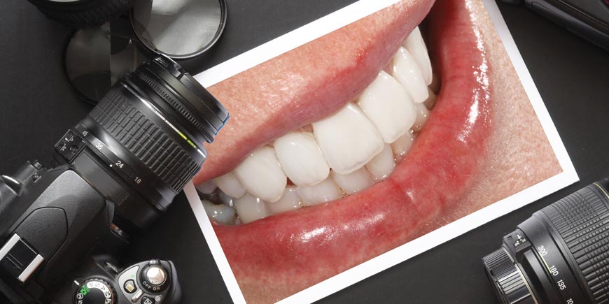
- Frontal at Rest It allows us to evaluate your facial features and characteristics, shape and proportions of the face; symmetry and diversity, volume and contour of the lips; among many other qualitative characteristics.
- Frontal with Exaggerated Smile Its main objective is to determine and evaluate the relationship between the lips, teeth and gums when smiling (smile line). The gummy smile, characterized by an exaggerated exposure of the gums, is a very unattractive situation from the aesthetic point of view and determines many of the cosmetic and restorative treatments that are dispensed in normal practice. Another important utility of this projection is to evaluate the coincidence of the dental midline with the facial midline, since in cases of severe discrepancy, it is pertinent to plan its correction.
- Right Profile Essential to determine the facial profile and the degree of lip protrusion. Profiles can be straight (ideal), concave or convex, and their correction can often be included in the overall treatment plan. Orthodontics, orthognathic surgery, plastic surgery and oral rehabilitation with fixed prosthesis; are just some of the procedures that will allow you to improve your profile and facial appearance.
- Left Profile It is the contralateral complement of the anterior projection.
- Frontal Dentogingival with Natural Smile It is essential to contrast it with the image of the maximum smile and to make decisive decisions in dental aesthetics and oral rehabilitation procedures. It should be a close-up photo showing only lips, teeth, and gums. Be on the lookout for your study, as it is often overlooked and not done.
2- Intraoral Digital Photographs
They allow us to know your oral condition, especially in those cases where it is not possible to make a direct clinical evaluation.
The set of these oral images must also include, at least, 5 images:
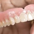
- Anterior or Frontal It shows the shape, texture, size and color of your front teeth. In addition, they are useful for detecting dental or gum pathologies. Caries, old or defective restorations, fractures, wear, gingival inflammation, exposed roots and gum abscesses; will never go unnoticed in an image of this type.
- Right Buccal Lateral photographs are used primarily to study the patient’s bite or occlusion. They allow us, among many other things, to appreciate how the teeth of both arches are related to each other when they come into functional contact, what is the type of dental intercuspation of the patient and what vertical space (interocclusal) we have to make the crowns, bridges or any other type of dental restoration. It is mandatory to take them with special rhodium or metal intraoral mirrors, to avoid double images, achieve a perpendicular projection of the surfaces and overcome the distortions inherent to oblique shots, which alter the occlusal reality.
- Left Buccal It is the contralateral complement of the anterior image.
- Maxillary Occlusal (Upper) It provides a broad view of the size and shape of the maxillary dental arch. It is used to determine the number of teeth present and absent, the dental arrangement and alignment, the length of the edentulous spaces and the transverse characteristics of the alveolar ridge; which is very important for the placement of dental implants. Like lateral projections, they must be taken with mirrors to achieve a perpendicular view of the occlusal surface.
- Mandibular Occlusal (Lower) It corresponds to the same anterior projection, but in the lower jaw or mandible.
If I Have Few or No Teeth in My Mouth, Are Photographs Also Necessary?
Of course!
In edentulous patients, candidates for dental implants, it is also necessary to know their facial characteristics, the relationship between the gums and the lips when smiling, the morphological characteristics of the maxillary bone, the sagittal relationships between the residual ridges and the clinical conditions of the oral mucosa.
A complete photographic study is always a mandatory routine in any major cosmetic or restorative treatment, and constitutes a fundamental element in the diagnosis and planning case process.
“Currently the Digital Image, the Hypertext Transfer Protocol and the Internet; Allow Perfectly Studying, Diagnosing Cases and Planning Treatments at Distance”.
DENTAL TIP
Send Us All Your Diagnostic Studies, Visit Caracas and Save a Lot of Money!
Globalization and dental tourism are two extraordinary tools to access first level Dentistry. Currently millions of people travel to other countries in search of opportunities and prices that allow them to face their dental treatments; and thus, recover your smile.
Wherever you are, generally in a few hours you will be able to arrive in Venezuela, move to a comfortable hotel and immediately start your oral rehabilitation process with dental implants, porcelain veneers and 100% metal-free ceramic structures; at just a fraction of the price you would have to pay in your country of residence.
It is almost certain that to travel or not to travel will mean the difference between satisfaction and resignation, or at best, between simply getting “something” or being able to access “the best option”. At DENTAL VIP we are very good hosts and excellent professionals.
Contact us and find out how traveling to Venezuela can improve your self-esteem and quality of life!