Table of Contents
Projectional radiography is the practice of producing two-dimensional images using X-ray radiation. X-rays are a form of energy that can travel through or be absorbed by solid objects. This energy is absorbed by dense objects, such as teeth and bones, and show up on a film or image as light-colored areas. X-rays pass through less dense objects, such as gums and cheeks, and appear as dark areas in the radiographs.
X-rays or radiographs (two terms that are used interchangeably to refer to the study result) show all dental providers a lot. They help us see the condition of your teeth, roots, jaw placement and facial bone composition. They also help us find and treat dental problems early in their development.
Like many film mediums, including photography and movie making, radiography has gradually been making the switch to digital from conventional film. Today digital sensors are used and not physical polyester plates or films to register the images. Particular benefits of digital radiography include improved comfort for the patient and less cranial exposure to radiation. Digital images are also easily stored on a computer and can be easily transferred through the internet and other digital media. Always opt for digital radiographs to facilitate the workflow on your case.
Dental X-rays require no special preparation. The only thing you will want to do is brush your teeth before your appointment at the selected oral diagnostic center. That creates a more hygienic environment for those working inside your mouth.
Although exist many different types of radiological studies in Dentistry, they all are concentrated in two large groups of radiographs: extraoral and intraoral.
What Are Extraoral Radiographs?
Like the first part of the name suggests, extraoral X-rays are made with the sensor outside the mouth. This type of X-ray still shows the teeth but can also provide important information about the jaw and skull. Think of these X-rays like the “big picture” of oral health, they are used to see how everything comes together.
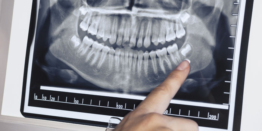
- Panoramic X-Ray Used to show the entire mouth on a single image. These pictures are used both for seeing tooth development and catching any emerging or impacted teeth, cysts and tumors.
- Cephalometric X-Ray Also called a ceph X-ray, these radiographs include the entire side of the head. It is used in both Dentistry and Medicine for orthodontic treatment planning, diagnosing issues such as sleep apnea and temporomandibular joint disorders.
- Cone-Beam Computed Tomography (CBCT) This type of X-ray is used for dental implant planning, evaluating the jaws and face, and much more. Like the name suggests, the images are taken using a cone-shaped X-ray beam that rotates around you taking three dimensional (3D) pictures.
- Standard Computed Tomography (CT) A standard CT scan may be done to determine size and placement location for implants. It does have a higher radiation exposure and must be done in a radiologist’s office or a hospital.
What Are Intraoral Radiographs?
Intraoral radiographs are the most common type of dental X-rays you will encounter during a routine dental exam. They are made with the sensor inside the mouth to detect cavities and verify the state of present teeth. These radiographs also give us the ability to view tooth roots, dental implants, check the health of the bone and even; diagnose periodontal disease. The different types of intraoral X-rays show different aspects of the teeth.
- Bitewing X-Rays These are done to help us locate decay or cavities between back teeth and bicuspids (teeth in front of the molars) that would be hard to find otherwise. The name comes from the wing-shaped device you bite down on while the X-ray is taken.
- Periapical X-Rays Focusing on two or three teeth, a periapical radiograph shows the entire length of the tooth, from crown to root.
- Occlusal X-Ray This X-ray (also known as palatal) shows the entire arch of teeth in either the top or bottom jaw. Since this type is larger than most intraoral X-rays it is often used to highlight tooth development and placement in children.
When Is a Panoramic X-Ray Necessary?
Absolutely in all cases, without exception!
The reality is that a panoramic X-ray is not only necessary to preserving your oral health, it could also save your life.
A panoramic is a two-dimensional radiograph that captures a picture of a patient’s entire mouth in just one image. The X-ray includes a view of all of the teeth, multiple bones of the head and neck, and other critical anatomical structures.
Unlike intraoral dental radiographs which show a close up of view of your teeth, this X-ray give us a global view of a patient’s head and neck. This view allows for diagnosing more than just regular dental concerns, like cavities or gum disease. Rather, this view enable us to see other important issues that may be occurring in the surrounding tissue and jaw bones like oral cancer or other abnormalities, things that would not be visible in an intraoral X-ray.
When Is an Intraoral Periapical Radiographic Study Indicated?
Intraoral periapical radiographs are widely used for diagnostic purposes due to its great precision, simple technique, low cost, less radiation exposure and widely available in clinical settings.
This type of radiography is indicated whenever the patient has natural teeth, dental implants or fixed restorations in his mouth; as a complement to panoramic radiography. However, when the patient does not have or has very few teeth left in the mouth, the single panoramic X-ray is usually sufficient to complete the oral diagnostic process.
Unlike panoramic radiography, periapical radiographs focus on the tooth and its supporting structures. The panoramic provides a greater field of vision (both full jaws), and the periapical one provides much closer and more detail (only 2 or 3 teeth per shot). With the panoramic view we see more and with the periapical one much better, that simple!
In addition to the aforementioned diagnostic objectives, periapical studies are also useful to assess the evolution of complex treatments.
How Do I Know if I Definitely Need or Not Diagnostic Periapical X-Rays?
It is always advisable to delegate that decision to your general Dentist.
However, if you do not have it, we recommend you do not worry and start from the idea that you need them. Once you are at the radiology center to take your extraoral radiographs (panoramic, Cone Beam tomography or both), the technician in charge will have enough professional judgment to tell you whether or not you need intraoral radiographs.
In the worst case, just send us your panoramic radiograph and we will promptly indicate if they are necessary or not. Remember that the only contraindication for this type of X-rays, apart from pregnancy, is the absence of teeth. If you still have more than 5, 6 or 7 teeth in your mouth, they will surely be necessary!
How Many Images Does a Periapical Study Include?
It depends on the quantity and arrangement of the teeth and restorations in the mouth. A full dentition (28-32 teeth) merits between 14 and 16 projections; however, the study could cover 1, 2, 3, 4, 5, 6, 7, 8, 9, 10, 11, 12, 13, 14, 15, or all 16 radiographs. There is no fixed number, it all depends on the case.
Generally, on average, a complete study includes the following fourteen X-rays:
- Eight Posterior Periapicals Two maxillary molar periapicals (left and right), two maxillary premolar periapicals (left and right), two mandibular molar periapicals (left and right), two mandibular premolar periapicals (left and right).
- Six Anterior Periapicals Two maxillary canine-lateral incisor periapicals (left and right), two mandibular canine-lateral incisor periapicals (left and right), two central incisor periapicals (maxillary and mandibular).
Full Mouth Series
It is simply a complete periapical study that also includes taking 2 bitewing X-rays, one right and one left. The full mouth series is composed of 16 or 18 films, taken the same day. Although bitewing radiographs are not essential, we always recommend including them in your study.
Why Should I Always Demand the Periapical Long Cone Paralleling Technique?
Because although there are many ways to take periapical radiographs, only the long cone paralleling technique is capable of generating accurate and reliable images.
This technique positions the sensor parallel to the long axis of the teeth and guides the central ray of the X-ray beam to be directed at a right angle to the teeth and the sensor. This method produces images of the teeth with minimal distortion.
A Rinn instrument is mandatorily used to help position and stabilize the film in the mouth as well as aim the X-ray beam. This device is comprised of a receptor holder/bite block, an aiming ring and a connecting rod.
Always demand the use of these devices and this technique to obtain quality radiographs and avoid unpleasant repetitions.
What Types of Problems Do Periapical Radiographs Help Detect?
Periapical X-rays help us diagnose with precision problems in your teeth and jaws. In adults, this type of radiographs show:
- Decay, especially small areas of decay between teeth.
- Decay beneath existing fillings.
- Bone loss in the jaw.
- Changes in the bone or root canals due to infection.
- Condition and position of teeth to help prepare for tooth implants, braces, prosthesis or other dental procedures.
- Abscesses (an infection at the root of a tooth or between the gum and a tooth).
- Cysts and some types of tumors.
“An Oral Diagnosis Without the Support of Radiographic Evidence, Will Always Leave Much to Be Desired”.
DENTAL TIP
Are Dental X-Rays Safe?
While dental X-rays do involve radiation, the exposed levels are so low that they are considered safe for children and adults. Because we always recommend an oral diagnostic center that uses digital X-rays instead of developing them on film, your risks of radiation exposure are even lower.
They will also place a lead “bib” over your chest, abdomen, and pelvic region to prevent any unnecessary radiation exposure to your vital organs. A thyroid collar may be used in the case of thyroid conditions. Children and women of childbearing age may also wear them along with the lead bib.
Pregnancy is an exception to the rule. Women who are pregnant or believe they may be pregnant, should avoid all types of X-rays. Tell us if you believe you are pregnant, because radiation is not considered safe for developing fetuses.
The Smile of Your Dreams Awaits You in Venezuela!
A cost saving alternative to your local dental care.
DENTAL VIP offers dental services in Caracas to patients from North America, Europe and the Caribbean.
We designed all our services to meet the special needs and expectations of our guests travelling for treatment:
- 50-70% lower prices.
- Volume discounts.
- Free consultation (X-ray, dental quote and transport).
- Extended guarantee packages for all treatments.
- Shorter treatment times and no waiting list.
- Free all time patient support.
- Travel and hotel assistance.
- After-care services.
We wish to ensure that your visit not only saves you money but you receive the best care for your dental needs. DENTAL VIP is dedicated to provide value for your money in your dental treatment, travel, and stay in Caracas.
Do not compromise on quality for the price, visit us in Caracas for your treatment and allow yourself the best. Do not settle for awkward removable dentures, low-end dental implants, or only temporary solutions.
Do not waste any more time, contact us right now!
