
Table of Contents
The temporomandibular joint (TMJ) is formed by the articulation of the mandible and the temporal bone of the cranium. It is located anteriorly to the tragus of the ear, on the lateral aspect of the face.
We all have two temporomandibular joints: one right and one left.
The TMJ is the only mobile joint of the face and whole head, and it is the one that allows, with the help of the facial muscles, that we can move the jaw to speak, eat and chew; among other functions.
Knowledge of these structures is essential in Dentistry, since the processes of dental occlusion and chewing depend on and are closely related to them.
In this post, we shall look at the anatomy of the temporomandibular joint, its articulating surfaces, ligaments and clinical correlations. In addition, we will know a little about the chewing muscles, which are what allow us to occlude (join) the teeth to cut, tear and grind food.
Articulating Surfaces
The temporomandibular joint is made up of three bony surfaces; the mandibular fossa and articular tubercle (both from the squamous portion of the temporal bone), and the condyle of the mandible.
This joint has a unique functional mechanism; the articular surfaces of the bones never come into contact with each other because they are separated by an articular disc. The presence of this disc divides the joint into two synovial cavities, each lined by a synovial membrane. The articular surface of the bones is covered by fibrocartilage and not by hyaline cartilage.
Ligaments
There are three extracapsular ligaments. They act to stabilize the entire temporomandibular joint.
- Lateral Ligament Runs from the beginning of the articular tubercle to the mandibular neck. It is a thickening of the joint capsule, and acts to prevent posterior dislocation of the joint.
- Sphenomandibular Ligament It is a band of fibrous tissue that joins the pterygoid processes of the sphenoid with the mandible on the inside.
- Stylomandibular Ligament A thickening of the fascia of the parotid gland. Along with the facial muscles, it supports the weight of the jaw.
Movements
Movements of this joint are produced by the muscles of mastication and the hyoid muscles. The two divisions of the temporomandibular joint have different functions:
Protrusion and Retraction
The upper part of the joint allows the jaw to protrude and retract, that is, its movements in an anterior and posterior direction.
The lateral pterygoid muscle is responsible for the protrusion (assisted by the internal pterygoid), and the posterior fibres of the temporalis the retraction. Lateral movements are achieved by antagonistic and contralateral protrusion and retraction actions of both joints (right and left).
Elevation and Depression
The lower part of the joint permits elevation and depression of the mandible; for opening and closing the mouth. Depression is mostly caused by gravity. However, if there is resistance, the digastric, geniohyoid, and mylohyoid muscles assist. Elevation is very strong movement, caused by the contraction of the temporalis, masseter, and internal pterygoid muscles.
Neurovascular Supply
The arterial supply of the TMJ is provided by the branches of the external carotid, principally the superficial temporal branch. Other contributing branches include the deep auricular, ascending pharyngeal and maxillary arteries.
The TMJ is innervated by the auriculotemporal and masseteric branches of the mandibular nerve (CN V3).
Muscles of Mastication
The muscles of mastication are a group of muscles made up of the temporalis, masseter, internal pterygoid and lateral pterygoid muscles. The temporalis muscle is situated in the temporal fossa, the masseter muscle in the cheek area, while the internal and lateral pterygoids lie in the infratemporal fossa.
As we already mentioned, the masticatory muscles are attached to the mandible, and are the ones that generate its movements, through the action of the temporomandibular joint (TMJ). These movements include:
- Protrusion (protraction), which moves the mandible forwards.
- Retraction, which pulls the mandible backwards.
- Elevation, which elevates the mandible and closes the mouth.
- Depression, which depresses the mandible and opens the mouth.
- Rotation, which produces side-to-side movements of the mandible.
All muscles of mastication are innervated by motor fibers of the mandibular branch of the trigeminal nerve (CN V3), while the arterial supply is mainly provided by branches of the maxillary artery.
Temporalis Muscle
The temporalis muscle is a large and flat muscle that lies within the temporal fossa of the skull. This fan-shaped muscle arises from the entirety of the fossa, below the temporal line, as well as from the deep surface of the temporal fascia. Its muscle fibers converge anteriorly to form a tendon that extends deep to the zygomatic arch. The tendon is then attached to the apex and medial surface of the coronoid process, and the anterior border of the ramus of the mandible.
The muscle is innervated by the deep temporal branches of the mandibular nerve, and vascularized by the deep temporal branches of the maxillary artery and middle temporal branches of the superficial temporal artery.
The temporalis muscle functions mainly as an elevator of the mandible. This function is largely produced by its anterior vertical fibers which are continually in action, opposing gravity when the mouth is closed. The contraction of the posterior and more horizontal fibers of the muscle, produces a retraction of the mandible, pulling it backwards. In addition, the temporalis muscle contributes to grinding movements, by moving the mandible from side to side.
Masseter Muscle
The masseter muscle is a strong, quadrangular muscle that covers the lateral aspect of the ramus of the mandible. It is composed of two layers that slightly differ in their attachments:
- Its superficial layer, bigger, arises from the maxillary process of the zygomatic bone and the anterior two thirds of the zygomatic arch. From their origin, these muscle fibers run down and back to join the lateral surface of the angle and the lower half of the ramus of the mandible.
- The deep layer of the masseter muscle arises from the medial surface and inferior margin of the zygomatic arch. These fibers run vertically downwards to insert onto the upper part of the ramus of the mandible and the coronoid process.
The innervation of the masseter muscle stems from the masseteric nerve, a branch of the mandibular nerve. Its blood supply is derived from the masseteric artery, which emerges from the maxillary artery. The major function of the masseter muscle is to elevate the mandible, with a minor contribution to the protrusion of the same.
Internal or Medial Pterygoid Muscle
The internal pterygoid muscle is a quadrangular muscle situated in the infratemporal fossa. It is composed of two heads that have two different sets of origins.
- The deep and bigger head arises from the medial surface of the lateral pterygoid plate of the sphenoid bone and the adjacent pyramidal process of palatine bone.
- The smaller superficial head originates from the tuberosity of the maxilla.
Both heads encircle the lower fibers of the external or lateral pterygoid muscle. From their points of origin, the muscle heads converge and run posterolaterally in an oblique fashion to insert on the medial surface of the mandibular ramus, close to the angle of the mandible.
The internal pterygoid muscle is innervated by the medial pterygoid branch of the mandibular nerve. Its principal blood supply stems from the pterygoid branches of the maxillary artery.
The major functions of this muscle are elevation of the mandible and side-to-side movements when grinding and chewing. The internal pterygoid is also involved in protrusion of the mandible.
External or Lateral Pterygoid Muscle
The lateral pterygoid muscle is a triangular muscle that lies in the infratemporal fossa. Like the internal pterygoid muscle, the lateral pterygoid has two heads, with two distinct origins.
- The smaller or superior head, that arises from the inferior surface of the greater wing and infratemporal crest of the sphenoid bone, which form the roof of the infratemporal fossa.
- The larger or lower head, originating from the lateral surface of the lateral pterygoid plate of the sphenoid bone.
The fibers of the two heads fuse and project posterolaterally to insert into a shallow depression in the anterior aspect of the neck of the mandible, called the pterygoid fovea. In addition, some fibers insert into the joint capsule and joint disc of the temporomandibular joint (TMJ).
The lateral pterygoid muscle is innervated by the lateral pterygoid branch of the mandibular nerve and vascularized by the pterygoid branches of the maxillary artery.
The function of the lateral pterygoid depends on the timing of its contraction. Bilateral contraction of the lateral pterygoid muscles protrudes and depresses the mandible. A unilateral contraction on a particular side, in conjunction with the ipsilateral medial pterygoid muscle, moves the mandible to the opposite side. This allows for alternating side-to-side movements during chewing.
“The TMJ Is the Only Mobile Joint of the Head and the One that Allows the Mandible to Be Moved to Speak, Eat and Chew”.
DENTAL TIP
Importance of TMJ in Dentistry
No Dentist will be truly competent if he does not know and master the anatomy and physiology of the temporomandibular joint and the masticatory muscles.
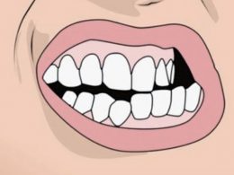
The teeth and gums are structures that act in synergy with muscles and joints through the phenomenon of dental occlusion, and therefore, they can never be approached and treated as isolated or independent units.
Your responsibility as a patient is to understand it and demand quality dental treatments that do not alter your normal occlusion and that allow you to chew comfortably and without any limitation. If you need extensive or complex restorative treatments, or feel discomfort when biting after prosthetic treatment, your best option is to go to a Specialist in Gnathology or Prosthodontics.
Making Dentistry Care Simple and Affordable…
DENTAL VIP is one of the most modern and recognized private dental clinics in Venezuela, and it could be your trustworthy partner in dental treatment abroad offering you: over 12 years of experience in dental tourism, excellent Dentists, high quality treatment covered by guarantee, comprehensive care, supportive and helpful staff, affordable prices and comfortable stay at our recommended hotels in Caracas.
All Dentists working at DENTAL VIP have a long experience in Dentistry and are highly qualified. Our Dentists, apart from having fourth level studies, regularly take part in domestic and foreign trainings, conferences and seminars.
The quality is guaranteed, and the price too! Do not waste any more time, contact us today and save up to 70% on extensive or highly complex dental treatments.