Table of Contents
Periodontal debridement is the procedure in which plaque and calculus (tartar or stone) that have accumulated on the teeth and gums, are mechanically removed. This is what we normally know as “dental cleaning”.
Root scaling and planing is a deep cleaning, below the surface of the gums, practiced to treat the disease of them. Scaling removes plaque and tartar that is inside the gums, while root planing softens the root of the tooth and helps the gums re-insert onto its surface.
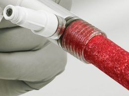
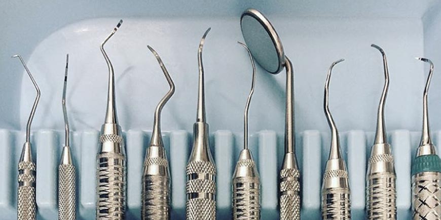
Debridement techniques include the use of hand tools, such as universal and Gracey curettes; as well as ultrasonic instruments, which fracture the calculus facilitating its removal.
Manual instrumentation has been the classically accepted technique for the demanding task of root debridement since the introduction of the curette in 1916. The appearance of the ultrasonic scaler in 1958 should have had a major impact on the profession, but achieve its widespread use was a slow and progressive process. Seven years after the original publications on these devices, many authors described the great benefits of using ultrasonic debridement during dental care. They were the first to document less patient fatigue and discomfort while undergoing periodontal treatment, as well as better visualization of the operative field.
In the early 1990s, ultrasonic instrumentation became an accepted modality of periodontal therapy, mainly due to an evolution in the size and shape of ultrasonic tips. Technological improvements in their design changed the perception of professionals about the use of ultrasound. Today, a wide variety of tips are available, from the thinnest ones used in subgingival instrumentation, to some special ones for dental implants.
Dental Plaque-Induced Gingivitis
Plaque-induced gingivitis is the most common form of periodontal disease seen by Dentists around the world.
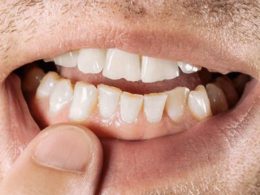
It is characterized by an inflammation of the gum as a result of the constant presence of a bacterial biofilm at the gingival margin. There may also be calculus. Although the clinical signs of gingivitis can vary between patients, the key factor that distinguishes it from periodontitis is the lack of attachment loss.
Probing depth should be modest, 2 or 3 mm, or slightly greater, due to gingival enlargement due to edema or hypertrophy. There should be no clinical or histological evidence of apical migration of the epithelial insertion. Radiographically, no bone loss should be seen.
Treatment Protocol for Gingivitis
The treatment protocol for this type of disease may begin with ultrasonic instrumentation or hand instrumentation.
If gross deposits exist, standard sized ultrasonic tips may be used. The tooth surfaces should then be evaluated with a manual instrument. An excellent choice is the number 11-12 explorer, a probe or a curette.
Mild to moderate residual deposits should be carefully removed using various curettes or fine ultrasonic tips. After removal of the deposit, a sickle scraper can be used to remove residual calculus or any interproximal staining. If the stain persists, the final steps may include polishing followed by a fluoride treatment.
Chronic Periodontitis
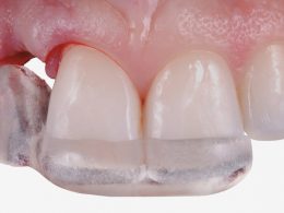
Chronic periodontitis is a disease related to dental plaque, like gingivitis, but it is characterized by the loss of the epithelial junction. The gingival tissues are usually red and inflamed, but if the patient forms fibrotic tissue, they may not be noticeable.
Chronic periodontitis was originally considered an age-related phenomenon, but it is now known to involve host resistance factors, in response to periodontal pathogens present in the mouth.
If the patient has more than 30% of his gums with loss of attachment, then the disease is considered to be generalized. Loss of fixation that affects less than 30% of teeth, is considered localized disease.
Also, the amount of attachment loss can, in a generic way, suggest the severity of periodontitis. An attachment loss of 4 mm suggests mild disease, 5 mm moderate disease and loss of 6 or more millimeters, suggests severe disease. However, the depth of the pocket or pathological sac cannot be used as the sole clinical sign to determine the extent of the process or severity of the disease, since certain conditions such as edema and hypertrophy can contribute to its depth, but in no case moment generate insertion loss.
Treatment Protocol for Periodontitis
The treatment protocol for patients with chronic periodontitis usually begins with ultrasonic instrumentation and standard-sized tips to remove any supragingival deposit. The next step should involve subgingival instrumentation, with thinner tips and used with sufficient power, depending on the characteristics of the deposit. When applying this type of instrumentation, the power setting is important, and it should always be the lowest one that allows the deposit to be removed.
After complete debridement with the thinner tips, the root surfaces should be evaluated and work continued with manual instrumentation, using area-specific curettes. The selection of the curette is based on the topography of the sac. Standard size curettes can be used in wide and deep pockets. Mini curettes should be used in narrow sacs. A fine-tipped explorer is helpful at this point to assess the texture of the root surface.
After final manual instrumentation for deposits removal, the thin ultrasonic tips can be used again in a low power setting to remove any stains or sludge from the root surface and smooth it out as much as possible. Diamond files can be used by hand as final finishing instrumentation, to achieve a smooth root surface free of any surface deposit or stain. In moderate or severe cases, an antimicrobial agent is usually applied.
Optional final steps include polishing the teeth and applying fluoride.
The more advanced the disease, the longer the initial treatment will take. An incipient case of chronic periodontitis may require only one to one and a half hours of instrumentation. An effective protocol for a moderate case is two visits, each lasting an hour and a half or two hours. The right side of the mouth can be completed in one visit, and then the left side in another. In a severe case, three two-hour visits or four hour-and-a-half visits may be necessary.
It is difficult, if not impossible, to establish absolute guidelines on how long a specific case may require to be well instrumented. Exist many variables, including the hardness of the deposit, difficulty of access to the root surfaces due to anatomical variables, the depth of the pocket, the presence of over-contoured restorations, the degree of gingival inflammation and the skill or dexterity of the professional; among other. The instrumentation must be prolonged until the root is completely free of deposits.
Severe chronic periodontitis is the most difficult to treat. Often, multiple teeth can be lost. Permanent and lifelong monitoring with a Periodontic Specialist is the best treatment plan for such a periodontal patient.
“The Main Objective of Cleanings and Root Scalings, Is to Remove Calculus and Smooth the Roots of the Teeth to Treat and Control Periodontal Disease”.
DENTAL TIP
A Blended Instrumentation Is Our Modern and Successful Approach to Periodontal Treatment!
It allows us the total elimination of supragingival deposits, without damaging the dental enamel. In more advanced cases, we use thinner tips for the deeper surfaces of the roots. These tips, applied with adequate power, can remove almost all of the subgingival calculus with little impact on the integrity of the root cement.
The next step in our protocol is manual instrumentation, which facilitates debridement at the level of grooves and anatomical accidents. Advances in the design of these instruments, allow us to achieve a better finish in all root scaling procedures.
To finish, we use ultrasound again, now with low power and ultrafine tips; to irrigate, remove already detached debris and smooth the root surface.
This combined instrumentation protocol can also be modified with the use of medications and other manual instruments, according to the exigencies of the case. Its ultimate goal is none other than to achieve effective treatment to improve the periodontal health of all our patients.
Save Up to 70% on Your Dental Quote!
Never forget that dental tourism is an extraordinary tool for accessing top-notch Dentistry.
DENTAL VIP trusts that quality of dental care should be affordable to all, no matter where you live. Hundreds of patients come to Venezuela for state-of-the-art medical and dental care every year, saving large amounts of money on health. Our rates are significantly lower than in any other Latin American country, without it implying a decrease or compromise in the quality of service. Simply what happens is that property costs and salary levels are both much lower than in any other place, which translates in better offers for our patients.
We guarantee that by using the services offered by DENTAL VIP, you could save up to 70% on your dental quote, compared to the local rates stipulated in your country.
Come for the price and stay for the quality! Contact us today via WhatsApp +58 414-9033547 or Email and overcome once and for all any barrier that prevents you from show off a white, healthy and beautiful smile.